
|
The major thrust of the 7-BM beamline is the application of synchrotron radiation tools to examine complex fluid flowfields. Three major techniques are applied: radiography, phase contrast imaging, and x-ray fluorescence spectroscopy. While optical techniques are often ideally suited to the study of fluid flowfields, there are certain flowfields for which optical diagnostics have significant challenges. These include:
|
||
| Radiography | ||
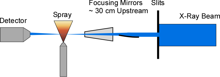
X-ray radiography makes use of the fact that at low to moderate x-ray energies (<10 keV), the dominant interaction of x-rays with materials is photoelectric absorption. As photoelectric absorption is generally governed by interactions with the core shell electrons, the absorption is relatively insensitive to physical phase or chemical bonding (except near absorption edges). As such, a simple Beer-Lambert law approach can be used to quantitatively determine the projected density of material in an x-ray beam. While radiography can be performed using laboratory x-ray sources, the use of a synchrotron source provides several advantages over laboratory sources. The overall flux from synchrotron sources is much greater than from laboratory sources. By creating a monochromatic beam, the synchrotron radiography measurements are no longer subject to beam hardening effects, which are present in measurements with polychromatic laboratory sources. The lack of beam hardening greatly simplifies data analysis. Finally, the collimated nature of synchrotron x-rays allows for efficient focusing of the x-rays. The high-intensity, monochromatic, focused beam available at 7-BM has been used to perform high-resolution, quantitative, time-resolved measurements of a wide variety of multiphase flowfields. These have included diesel injection, gasoline injection, cavitation, aerated liquid sprays, and rocket injection.
|
||
| X-Ray Fluorescence Spectroscopy | ||
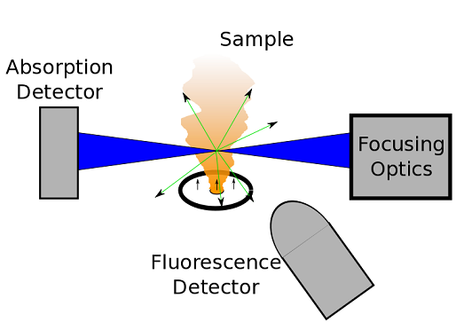
In addition to radiography, 7-BM is also equipped with a silicon drift diode detector for performing x-ray fluorescence spectroscopy. In x-ray fluorescence, an atom emits an x-ray photon after absorbing an incident x-ray photon. The energy of the emitted x-ray photon depends on the element of the atom which emitted the photon. As such, fluorescence spectroscopy is widely used for nondestructive elemental analysis. Fluorescence has distinct advantages over radiography in certain flowfields, including element specificity, relative insensitivity to changes in beam intensity, and greater flexibility in energy range. The maximum time resolution attainable with fluorescence is significantly lower than that with radiography. |
