|
1-ID techniques leverage the high-brilliance of synchrotron x-rays in the 42<E<120 keV range to non-destructively probe bulk materials. Often the techniques listed below can be combined on a single specimen, to obtain multi-modal data that span a wide range of length scales. These techniques can also be conducted in situ using external stimuli including temperature and mechanical loading.  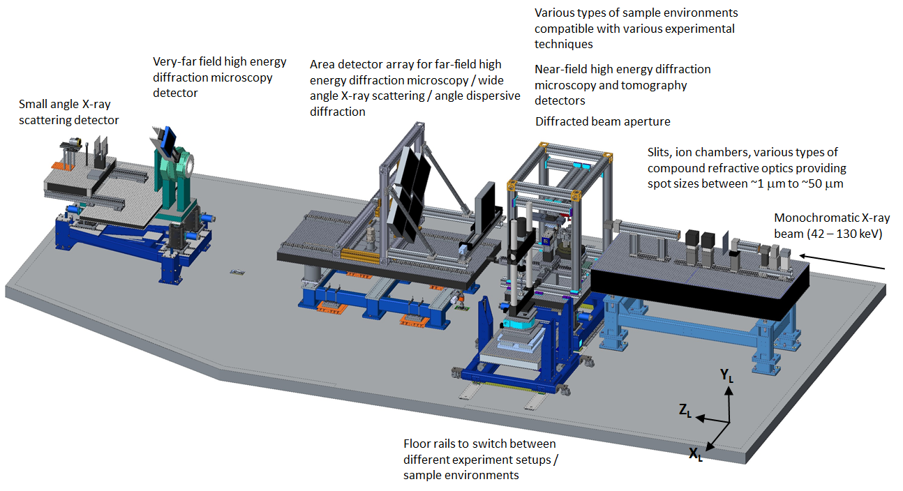 |
||||||||||||||||||||||||||||||||||||||||||||||||||||||||||||||||||||||||||
At 1-ID, Wide Angle X-ray Scattering (WAXS) technique employs a transmission geometry and large area detector(s) to measure grain-averaged quantities such as phase fraction, internal strain, dislocation density and crystallographic texture in each phase of a polycrystalline material. Such materials encompass the vast majority of structural materials, ranging from aircraft wings to engine crankshafts to bones and teeth. The high-flux of synchrotron x-rays combined with modern area detectors allows for weak phases to be detected, such as Zr-H phases in Zr alloys with H concentrations on the 100 parts-per-million level, and rapid data collection with less than 1 s/detector frame common. At the low Bragg angles associated with these high-energies (2θ(typ) < 15 degrees), the area detector captures diffraction information from a 2D plane of sample orientations which are nearly perpendicular to the incident beam. The incident x-ray beam size can be decreased to approximately 2 (V) x 10 (H) microns using slits and/or focusing elements, allowing gradients transverse to the incident beam to be probed by translating the sample relative to the fixed beam. In the longitudinal direction, diffraction is typically averaged over the entire sample thickness traversed by the x-ray beam; however diffracted beam apertures may be used to produce longitudinal resolutions down to ~100 microns. By taking diffraction patterns as a function of a rotation, about an axis perpendicular to the incident beam (ω), multi-dimensional quantities may be measured including the orientation distribution function (macro-texture) and full strain tensor. In addition, this rotation provides 3D spatially-resolved WAXS reconstructions via the high-energy diffraction microscopy (HEDM) and scattering tomography (ST) techniques. Examples
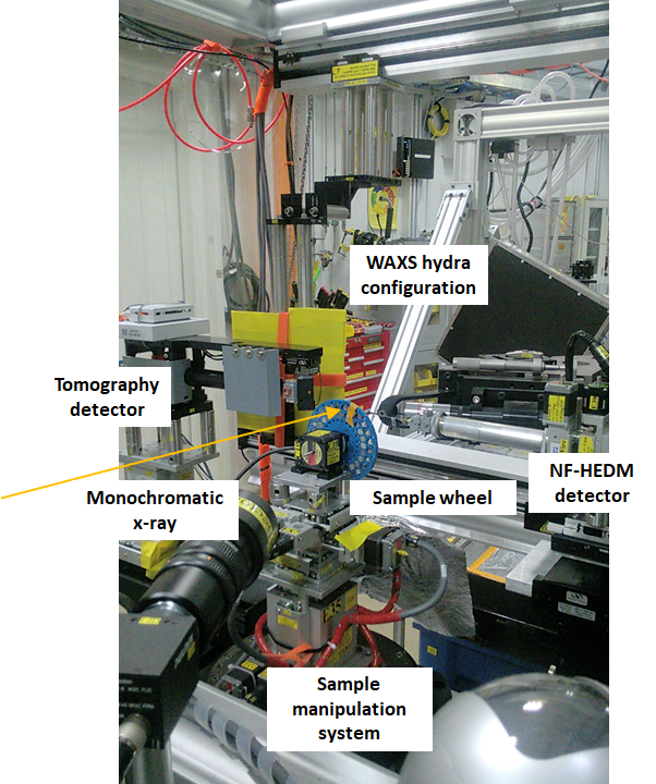
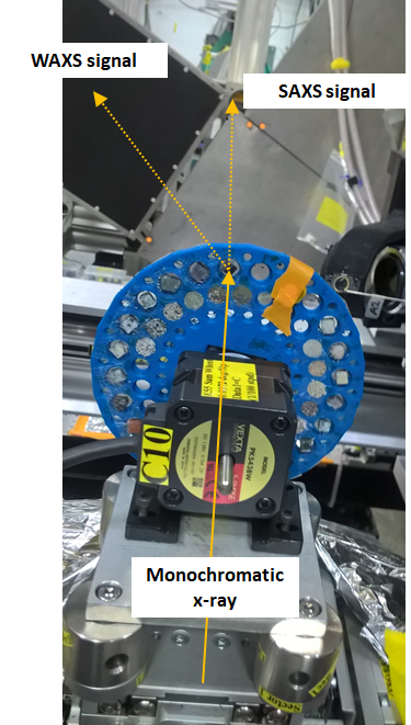
Small Angle X-ray Scattering (SAXS) is a pinhole-based technique which uses an area detector and ~6m sample-detector distance to determine the distribution of nanoparticle or void sizes as well as periodicity of long-range orders in hierarchical materials. SAXS is often combined with WAXS and the two signals can be captured concurrently using strategically located detectors as illustrated in the panoramic view of the 1-ID-E hutch. Examples
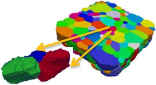
In the HEDM technique, individual grains within a polycrystalline aggregate can be mapped by taking diffraction patterns as a function of rotation angle. HEDM requires that diffraction spots from individual grains can be identified, meaning that there is an upper limit to the number of grains which can be measured in a given diffraction volume. In non-deformed samples as many as several thousands of grains may be indexed, while this number decreases with crystallite disorder (e.g. plastic deformation). There are 2 flavors of HEDM: near-field and far-field. In near-field HEDM the detector is placed several mm from the sample and sample-orientation is mapped with high (micron level) spatial resolution. In far-field HEDM the detector is about 1m from the sample and grain-level quantities are mapped with high q-resolution. Taken together, these techniques produce maps of crystallographic parameters in the illuminated volume including lattice parameters, orientations, positions and volumes and grain shape. Currently only a few synchrotron facilities are available worldwide which can generate this type of diffraction data. The closest non-x-ray analogue is electron backscatter diffraction, available much more widely, which provides 2D (surface) information with higher spatial resolution but poorer resolution of crystallographic parameters. Examples
The Scattering Tomography technique is based on collecting position-resolved 'powder-like' WAXS data at a number of angular orientations of the specimen, with positional intervals generally matching the beam-size and number chosen to cover the entire specimen cross-section.Through subsequent reconstruction of this data, the cross-section is partitioned, or voxellated, such that powder diffraction data can be attributed to each voxel.Three-dimensional imaging can be performed by an orthogonal specimen translation and repeating the process. This method is used for 3D volume reconstructions in lieu of HEDM when grain sizes are too small to be individually resolved with HEDM (roughly this limit is in the 1-5 micron range), and thus valuable for studying inhomogeneities in nano-grained materials including bio-materials and many energy-storage materials. High-energy monochromator characteristics Available beam sizes
WAXS detector
SAXS detector
Tomography detector
NF-HEDM detector
Detector distances
Typical calibration samples used
The APS Upgrade project (APS-U) will be an exciting endeavor that will boost the coherence and brilliance of the x-ray beam by replacing the existing APS storage ring and associated photon delivery infrastructure. To take advantage of the new highly coherent and brilliant beam, 1-ID will also undergo various enhancements so that we can continue to deliver world-class in situ experimental capabilities after APS-U. One of the new experimental techniques that is anticipated to come on-line is Bragg Coherent Diffraction Imaging (BCDI). The BCDI technique has been demonstrated at lower energy x-rays and utilized to measure the intra-granular structural changes in single, often isolated, particles.
In preparation for APS-U, 1-ID has been collaborating with ANL-Material Science Division's Synchrotron Studies of Materials group (https://www.anl.gov/msd/synchrotron-studies-of-materials) to develop the experimental and analysis infrastructure for BCDI at high-energy x-rays and combining it with HEDM to broaden the length scales that our techniques can cover. |
||||||||||||||||||||||||||||||||||||||||||||||||||||||||||||||||||||||||||