
| Multiple photon ionization |
|
The AMO group performed some of the first experiments on intense x-ray interactions with atoms and molecules using the LCLS x-ray free-electron laser (XFEL). A dramatic early observation was the stripping of all ten electrons from atomic neon by single LCLS pulses. The process of sequential absorption of six photons and four Auger decays is illustrated in Fig. 2. The experiments also demonstrated efficient hollow-neon production by sequential photoionization of both K-shell electrons prior to Auger decay. In another experiment on atomic neon, a single x-ray pulse first ejected a valence electron and then excited a K-shell electron to the valence hole. This was the first example of using intense x rays to open and excite a ``hidden resonance,'' which is a technique we are exploiting in studies of molecular core-hole decay dynamics. 
The capabilities of the LCLS and other XFELs are continually improving with increased control of pulse durations, reduced bandwidths and increased coherence by seeding, and schemes to produce two or more pulses with variable delays and colors. Our recent experiments seek to exploit these properties for studies of multiple-photon ionization and core-hole decay dynamics. |
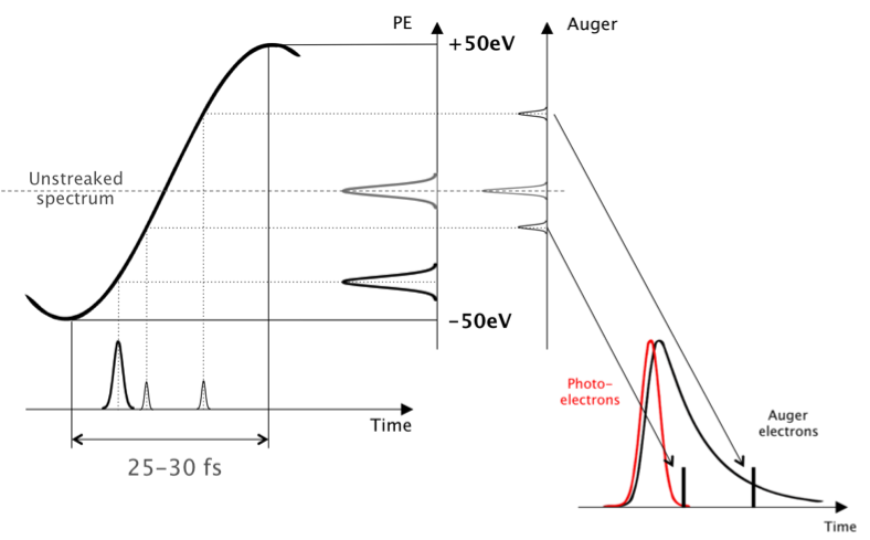
| LCLS pulse duration measurements |
The main route followed by our collaboration to get a handle on those temporal properties consists in transferring the time properties of the x-ray pulses to electron wave-packets produced during ionization or subsequent Auger decay of a gas target. The simultaneous presence of a strong laser field (operating in the visible, IR or THz region) modifies the energy spectrum of those electron wave-packets in a deterministic way, as shown in Fig. 3. The laser field "streaks" the electron energies as a function of the time of emission so that an electron energy measurement encodes time information. |
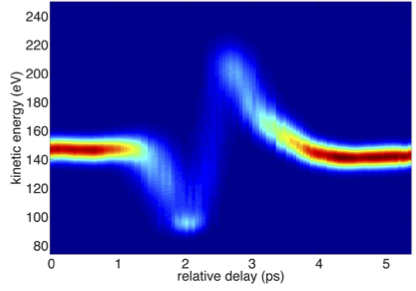
| Self-referencing time domain measurements of femtosecond inner shell dynamics |
Measuring the photoionization event allows one to create, on a shot by shot basis, an absolute reference for the timeline of events being triggered by that pulse. Because this timing is extracted for every shot, events with low statistics can then be correctly added up to yield high quality data in the time domain. The objective is to measure, for every shot, the streaked energy spectra of both the photoelectron and the other electrons of interest. A streaked photoelectron spectrum collected with good statistics allows determining, for a particular shot, the relative position inside the streaking ramp. Then, determining the amount of streaking experienced by the other electrons that are detected can directly indicate their emission time inside the ramp and relative to the x-ray pulse. |
| Hetero-site-specific femtosecond-time-resolved dynamics |
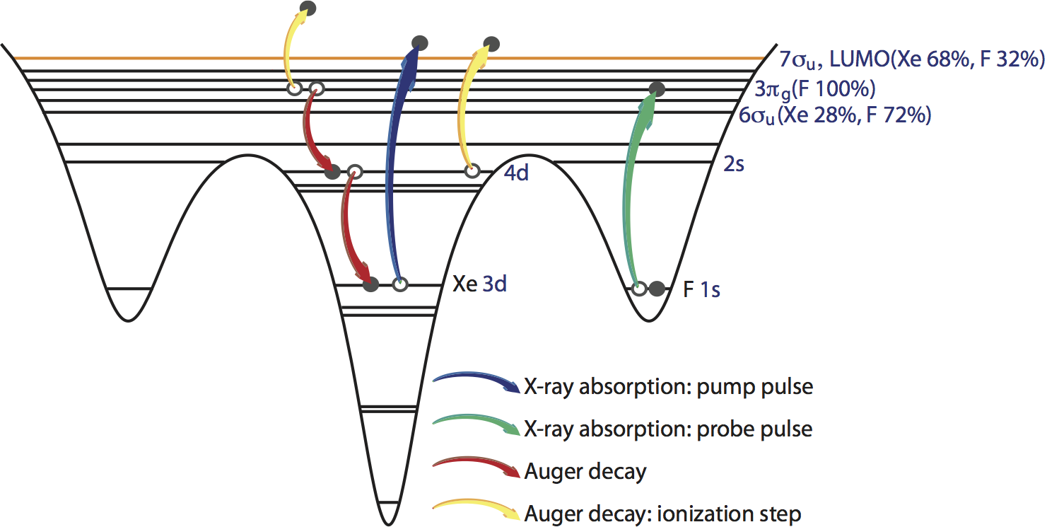
|
| Radiation damage in nanoparticles by intense x-ray interactions |
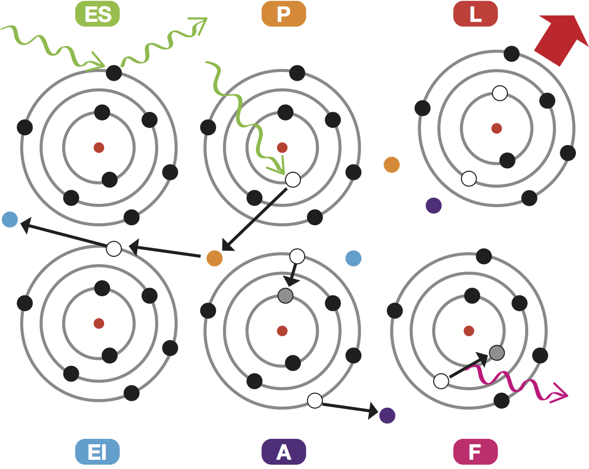
In order to obtain an atomistic view of the dynamical x-ray damage processes and the subsequent structural distortion on the target system throughout the x-ray pulse, we use a combined Monte-Carlo/Molecular-dynamics (MC/MD) computational model (see Fig. 6). The rates of all inner-shell transitions and the cross sections of photoionization of each subshell are obtained with Hartree-Fock-Slater calculations. During the x-ray pulse, the occurrences of the photoionization, inner-shell decay processes, electron-impact ionization and the site of their occurrences are treated by Monte-Carlo type methods. Subsequently, the dynamics of the photoelectrons, Auger electrons, secondary electrons and the atoms/ions in the systems are tracked using molecular dynamics methods. The advantage of this MC/MD model is that it allows us to compute the time-dependent coherent x-ray diffraction pattern of the target system by tracking the position of the delocalized electrons and the configuration (electronic configuration and charge state) and positions of atoms/ions. In addition, we can monitor the fluorescence spectrum, charge-state distribution, energy spectra, lattice dynamics, and dynamics of photoelectrons, Auger and secondary electrons. |