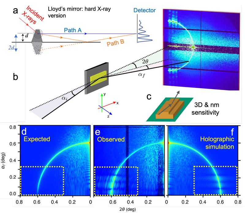
One of the most powerful and versatile imaging techniques developed in recent years for materials and biological science is X-ray coherent diffraction imaging, a lensless technique that can probe nanoscale and mesoscopic structures using diffraction patterns from highly coherent X-ray beams. Diffraction imaging has been commonly performed in transmission geometry where samples weakly perturb the incident X-rays, with reconstruction done using algorithms based on well-developed Fourier transform techniques.
However, reconstructing three-dimensional surface or interfacial structures supported by thick substrates has proved daunting due to strong absorption of the substrates and the requirement of reflection geometry. Strong reflective scattering from the supporting substrates or interfaces can render conventional reconstruction ineffective, requiring new analytical approaches.
Researchers at Argonne National Laboratory, working with collaborators from DESY in Germany, took inspiration from a classic physics experiment to develop a new technique for obtaining precise 3D structural information in only a single view. Their work appeared in Nature Communications.
English polymath Thomas Young demonstrated how light from two different sources could create distinctive interference patterns, pointing to the wave nature of light in his famous double-slit experiment. Humphrey Lloyd extended Young's work by using a mirror to create interference patterns between a single light source and its reflection, an experiment known as "Lloyd's mirror." Such intriguing phenomena, however, are far more difficult to demonstrate at the extremely short wavelengths of hard X-rays, thousands of times shorter than visible light.
In their current work, the investigators realized that the holographic patterns seen in hard X-ray grazing-incidence reflection geometry are an analog for a more conventional Lloyd's mirror setup to provide data for high-resolution 3D reconstructions of surface and interfacial structure. The team used both simulations and imaging experiments at the 8-ID-I beamline of the Advanced Photon Source (Argonne National Laboratory) and the P-10 beamline of PETRA-III (DESY Lab) to develop techniques for extracting 3D profiles of planar surface patterns at nanometer and sub-nanometer resolution.
The great complexity and unpredictability in surface scattering and speckling patterns from the grazing-incidence X-ray probes, which can also include substrate and interfacial reflections, make the interpretation of morphological information quite challenging. One approach for dealing with these problems is known as distorted-wave Born approximation (DWBA), a strategy that the Argonne investigators adapted and extended in a new approach they call finite-element DWBA (FE-DWBA). Using a 3D grid system, they were able to accurately compute X-ray scattering from a variety of heterogeneous features to generate high-resolution holographic 3D structural imaging.
To confirm and validate this approach, the experimenters conducted extensive simulation and experimental studies. They first studied an ultrathin gold bar on a silicon substrate, designed in a simple configuration so that its features could be derived from tell-tale scattering information. However, they found that strong in-plane inhomogeneities, along with the local electron densities far different from the averaged ones at the sample surface and substrate, compromised the efficacy of the usual DWBA approach for revealing structural features.
The research team addressed that problem with their FE-DWBA technique, in which the sample is considered not only as merely a layered structure but divided into a 3D grid of parallel and perpendicular sections, which allows computations to be done on the basis of the 3D unit cells. The resulting computational demands of dealing with up to five million cells in a single sample can be managed by grouping cells into stacks with the same electron profile and employing a first-principles simulation. Additional holography experiments on a sample with a more complicated and truly 3D configuration, using two stacked gold bars of differing widths, provided further confirmation of the holographic Lloyd's mirror effect.
The extreme sensitivity and nanometer resolution of the 3D imaging that can be obtained using this "optics-on-a-chip" approach offer immediate benefits for semiconductor metrology, providing a means of nondestructive visualization of nanoscale circuit configurations and patterns without any modifications to the devices. The researchers envision further development of this approach to achieve single-view, in situ and operando characterization in a variety of other research fields in both the physical and biological sciences. The scientists are continuing to expand upon and extend their work to extend its capabilities to full real-space 3D reconstruction imaging. – Mark Wolverton
____________________________________________________________________________________
See: M. Chu1,, Z. Jiang1, M. Wojcik1, T. Sun1,2, M. Sprung3, J. Wang1, “Probing three-dimensional mesoscopic interfacial structures in a single view using multibeam X-ray coherent surface scattering and holography imaging,” Nature Communications 14 5795 (September 2023).
Author affiliations: 1Argonne National Laboratory; 2University of Virginia; 3Deutsches Elektronen-Synchrotron (DESY).
We acknowledge DESY (Hamburg, Germany), a member of the Helmholtz Association HGF, for providing experimental facilities. Parts of this research were carried out at PETRA III, and we would like to thank Fabian Westermeier and Dmitry Dzhigaev for their assistance at the P10 beamline. Beamtime was allocated for proposal I-20170155. We acknowledge the fruitful discussion with Sunil K. Sinha. We also thank Wei Jiang for our early discussion on the algorithm and Suresh Narayanan and Raymond Ziegler for their valuable assistance with the experiments at Sector 8 of the Advanced Photon Source (APS). We acknowledged that Jong Woo Kim and Pice Chen who contributed to the sample preparation and the data collection and Donald Walko who read and commented on part of the manuscript. Work was also performed at the APS Center for Nanoscale Materials of Argonne National Laboratory (ANL), both US Department of Energy (DOE) Office of Science (SC) User Facilities. J.W., Z.J., M.C., M.W., and T.S. acknowledge the funding support provided by Laboratory Directed Research and Development (LDRD, 2017-073-N0) from ANL and by the US DOE, SC, Office of Basic Energy Sciences (BES), under Contract No. DE-AC02-06CH11357. J.W., Z.J., and M.C. acknowledge partial funding support from the Accelerator and Detector Research Program of the US DOE, SC, BES under Contract No. DE-AC02-06CH11357. Z.J. acknowledges partial support from the US DOE Early Career Research Award.
The U.S. Department of Energy's APS at Argonne National Laboratory is one of the world’s most productive x-ray light source facilities. Each year, the APS provides high-brightness x-ray beams to a diverse community of more than 5,000 researchers in materials science, chemistry, condensed matter physics, the life and environmental sciences, and applied research. Researchers using the APS produce over 2,000 publications each year detailing .impactful discoveries and solve more vital biological protein structures than users of any other x-ray light source research facility. APS x-rays are ideally suited for explorations of materials and biological structures; elemental distribution; chemical, magnetic, electronic states; and a wide range of technologically important engineering systems from batteries to fuel injector sprays, all of which are the foundations of our nation’s economic, technological, and physical well-being.
Argonne National Laboratory seeks solutions to pressing national problems in science and technology. The nation's first national laboratory, Argonne conducts leading-edge basic and applied scientific research in virtually every scientific discipline. Argonne researchers work closely with researchers from hundreds of companies, universities, and federal, state and municipal agencies to help them solve their specific problems, advance America's scientific leadership and prepare the nation for a better future. With employees from more than 60 nations, Argonne is managed by UChicago Argonne, LLC, for the U.S. DOE Office of Science.
The U.S. Department of Energy's Office of Science is the single largest supporter of basic research in the physical sciences in the United States and is working to address some of the most pressing challenges of our time. For more information, visit the Office of Science website.
