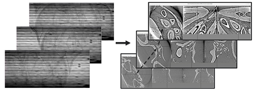
It is one thing to study crystals, battery cathode materials, or other types of inorganic samples using tomographic x-ray microscopy (and the technique has become invaluable for such investigations). But the study of biological samples poses a new range of complex challenges. Unlike inorganic materials, which are generally uniform throughout, organic samples are often one of a kind and irreplaceable, making any kind of destructive experiment preparation highly undesirable. They can also be sensitive to damage from strong doses of x-rays that can distort or destroy the structures under study. Tomographic imaging of large samples combining many different regions of interest also generates huge volumes of data that are computationally very expensive and time-consuming to reconstruct. To address these issues, a trio of researchers from Argonne National Laboratory has proposed an instrument for three-dimensional (3-D) real-time imaging at the U.S. Department of Energy’s Advanced Photon Source (APS) that can non-destructively create tomographic reconstructions of three separate slices of a biological sample and select certain specified regions for high-resolution scanning. Their work was published in the journal Microscopy and Microanalysis.
The investigators demonstrated the challenges presented by biological samples using as an example a tomographic study of a feline spinal cord. A sample measuring 6.9 x 6 x 2.9 mm3 was scanned with a resolution of 0.69 µm, in a mosaic of 5 x 2 overlapping tiles. Each tile contained 6000 projection angles at 1.9 second exposure each. The scan took 31.7 hours total and yielded 324 gigabytes of 16-bit data. The full tomographic reconstruction amounted to 1.1 terrabytes of 32-bit data.
Obviously, this is a complicated and time-intensive procedure, involving full 3-D reconstruction of the entire data volume. The instrument proposed by the researchers offers a far more efficient approach that permits the selection, saving, and reconstruction of only the most relevant data from three representative slices, which can be selected in real time. To demonstrate the technique, they examined a large beetle sample with reconstruction of three regions at the APS X-ray Science Division Imaging Group’s 2-BM beamline. The x-ray tomography instrument was integrated with an Optique Peter microscope to allow real-time zooming to regions of interest; this instrument also features an automatic lens changer for different magnifications. Data are processed in real time via two separate pipelines, one to stream projection data to a graphics processing unit for reconstruction, and the other to handle on-demand capture of selected data in Hierarchical Data Format version 5 files.
With low-resolution images obtained at 1.1 x magnification, and high-resolution at 5 x magnification, zooming to regions of interest of the sample is accomplished simply by moving the rotation stage using the x, y, and z motors of the sample stack. Scanning is performed with exposures of 0.05 s at 1.1 x with 360 projection angles and 0.1 second at 5 x with 720 projection angles. Parameters such as exposure time, rotation speed, and step size can be changed at any time as data streaming proceeds.
This system significantly reduces data size and acquisition time by allowing only the most essential images, chosen quickly and easily in real time, for the data-intensive process of full reconstruction. Because of the fast scanning, speed the radiation dosage to the sample is also sharply decreased, which lessens potential damage. The real-time nature of the technique proposed by the investigators could also allow in situ of some processes under changing conditions, again in real time (Fig. 1).
In this work, the Argonne researchers offer a fresh, more efficient, and highly versatile alternative to conventional approaches to tomographic -ray biological imaging. By saving the always-precious resources of image acquisition time and computer processing, this approach can preserve them for other imaging tasks that definitely require the maximum available power for both. ― Mark Wolverton
See: Viktor Nikitin*, Pavel Shevchenko, and Francesco De Carlo, “Real-time 3D Tomographic Imaging of Biological Samples with Zooming to Features of Interest,” Microsc. Microanal. 28 (Suppl 1), 1536 (2022). DOI: 10.1017/S1431927622006171
Author affiliation: Argonne National Laboratory
Correspondence: * [email protected]
This research used resources of the Advanced Photon Source, a U.S. Department of Energy (DOE) Office of Science User Facility at Argonne National Laboratory and is based on research supported by the U.S. DOE Office of Science-Basic Energy Sciences, under Contract No. DE-AC02-06CH11357.
The U.S. Department of Energy's APS at Argonne National Laboratory is one of the world’s most productive x-ray light source facilities. Each year, the APS provides high-brightness x-ray beams to a diverse community of more than 5,000 researchers in materials science, chemistry, condensed matter physics, the life and environmental sciences, and applied research. Researchers using the APS produce over 2,000 publications each year detailing impactful discoveries, and solve more vital biological protein structures than users of any other x-ray light source research facility. APS x-rays are ideally suited for explorations of materials and biological structures; elemental distribution; chemical, magnetic, electronic states; and a wide range of technologically important engineering systems from batteries to fuel injector sprays, all of which are the foundations of our nation’s economic, technological, and physical well-being.
Argonne National Laboratory seeks solutions to pressing national problems in science and technology. The nation's first national laboratory, Argonne conducts leading-edge basic and applied scientific research in virtually every scientific discipline. Argonne researchers work closely with researchers from hundreds of companies, universities, and federal, state and municipal agencies to help them solve their specific problems, advance America's scientific leadership and prepare the nation for a better future. With employees from more than 60 nations, Argonne is managed by UChicago Argonne, LLC, for the U.S. DOE Office of Science.
The U.S. Department of Energy's Office of Science is the single largest supporter of basic research in the physical sciences in the United States and is working to address some of the most pressing challenges of our time. For more information, visit the Office of Science website.
