
The original Carnegie Mellon University press release can be read here.
The dimer structure of an important tumor-suppressing protein, phosphatase and tensin homolog (PTEN), the second most frequently mutated protein found in human cancer, has been established by an international group of researchers carrying out studies at the U.S. Department of Energy’s Advanced Photon Source (APS), an Office of Science user facility. Their findings provide new insights into how the protein regulates cell growth and how mutations in the gene that encodes the protein can lead to cancer. The study was published online in Structure, and will appear in the October 6, 2015 issue.
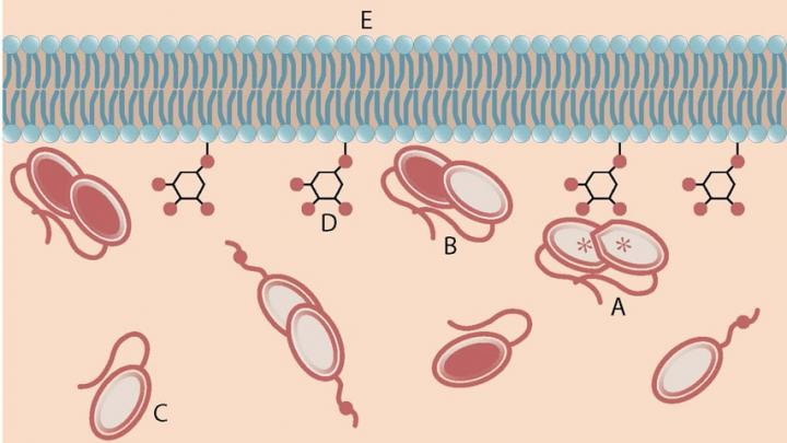
Phosphatase and tensin homolog is a known tumor suppressing protein that is encoded by the PTEN gene. When expressed normally, the protein acts as an enzyme at the cell membrane, instigating a complex biochemical reaction that regulates the cell cycle and prevents cells from growing or dividing in an unregulated fashion. Each cell in the body contains two copies of the PTEN gene, one inherited from each parent. When there is a mutation in one or both of the PTEN genes, it interferes with the protein's enzymatic activity and, as a result inhibits its tumor suppressing ability.
Membrane-incorporated and membrane-associated proteins like PTEN make up one-third of all proteins in our body; many important functions in health and disease depend on their proper functioning. Despite PTEN's importance in human physiology and disease, there is a critical lack of understanding of the complex mechanisms that govern its activity.
Recently, researchers at Harvard Medical School found that PTEN's tumor suppressing activity becomes elevated when two copies of the protein bind together, forming a dimeric protein. PTEN dimerization may be the key to understanding an individual's susceptibility for PTEN-sensitive tumors
In order to reveal how dimerization improves PTEN's ability to thwart tumor development, the researchers in this study, from Carnegie Mellon University, the National Institute of Standards and Technology, the Center for Synchrotron Radiation Research and Instrumentation, the Illinois Institute of Technology, Monash University, Harvard Medical School, the University of Massachusetts Medical School, and Worcester Polytechnic Institute needed to establish the protein's dimeric structure. Normally, protein structure is identified using crystallography, but attempts to crystallize the PTEN dimer had failed.
So the team used a different technique called small-angle x-ray scattering (SAXS), which gains information about a protein's structure by scattering x-rays through a solution containing the protein. This experimentation was carried out at the Biophysics Collaborative Access Team 18-ID-D x-ray beamline at the Argonne APS. They then used computer modeling to establish the dimer's structure.
They found that in the PTEN dimers, the C-terminal tails of the two proteins may bind the protein bodies in a cross-wise fashion, which makes them more stable. As a result, they can more efficiently interact with the cell membrane, regulate cell growth and suppress tumor formation.
Now that more is known about the structure of the PTEN dimer, researchers will be able to use molecular biology tools to investigate the atomic-scale mechanisms of tumor formation facilitated by PTEN mutations. The researchers also hope that their findings will offer up a new avenue for cancer therapeutics.
See: Frank Heinrich1,2, Srinivas Chakravarthy3,4, Hirsh Nanda1,2, Antonella Papa5,6, Pier Paolo Pandolfi6, Alonzo H. Ross7, Rakesh K. Harishchandra8, Arne Gericke8, and Mathias Lösche1,2*, “The PTEN Tumor Suppressor Forms Homodimers in Solution,” Structure (2015), in press, published on-line in advance of print. DOI: 10.1016/j.str.2015.07.012
Author affiliations: 1Carnegie Mellon University, 2National Institute of Standards and Technology, 3Center for Synchrotron Radiation Research and Instrumentation, 4Illinois Institute of Technology, 5Monash University, 6Harvard Medical School, 7University of Massachusetts Medical School, 8Worcester Polytechnic Institute
Correspondence: * [email protected]
This research was supported by the Department of Commerce (MSE 70NANB11H8139 and 70NANB13H009), NIGMS (R01 GM101647), NINDS (R01 NS021716) and the NSF (CHEM 1216827). This research used resources of the Advanced Photon Source, a U.S. Department of Energy (DOE) Office of Science user facility operated for the DOE Office of Science by Argonne National Laboratory under contract no. DE-AC02-06CH11357.
Argonne National Laboratory is supported by the Office of Science of the U.S. Department of Energy. The Office of Science is the single largest supporter of basic research in the physical sciences in the United States, and is working to address some of the most pressing challenges of our time. For more information, please visit science.energy.gov.