
 Light-oxygen-voltage (LOV) receptors are modular sensory proteins used by many organisms to perceive light. The blue light sensing apparatus is conserved among LOV proteins but the downstream response to light can vary depending on how the initial signal is transmitted to an effector module. For example, these proteins are known to activate movements toward light, regulate stress responses, and control DNA binding. The modular arrangement of LOV receptors has worked well to adapt light responses to the different needs of various species and has attracted interest in the world of bioengineering for the possibilities surrounding the synthesis of light-sensitive proteins that perform functions useful to humans. As a case in point, various LOV-based systems now allow researchers to use blue light to deliberately control a number of intracellular processes, including cytoskeletal dynamics and the subcellular location of proteins of interest. Although researchers have uncovered the photochemical mechanism underlying the LOV receptor light response, there is still a gap in our understanding of how the initial signal is coupled to activation of the downstream function. Now, new work on a homodimeric bacterial LOV called YtvA, carried out at the U.S. Department of Energy’s Advanced Photon Source (APS) has shed some light on this problem. The findings improve our understanding of how LOV transmits a signal from light to the associated effector protein that controls the response and opens the door to the development of bioengineered proteins that are responsive to light.
The research team in this study was a collaboration between scientists at the University of Gothenburg (Sweden) Humboldt University and Bayreuth University (Germany), the European Synchrotron Facility (France), the Paul Sherrer Institute (Switzerland), and The University of Chicago. The structure of the LOV domain from YtvA had been previously solved by x-ray crystallography, but the differences between the dark and light-activated structures of the protein were not large enough to conclusively point to a mechanism by which the structural changes would transmit a signal to the effector domain of the protein. Also, the structure was missing a helical domain that the team felt might be important to the activity of the LOV domain. They decided to do x-ray scattering experiments in solution with a form of the protein that contained that domain to see if they could observe structural changes that might provide more information.
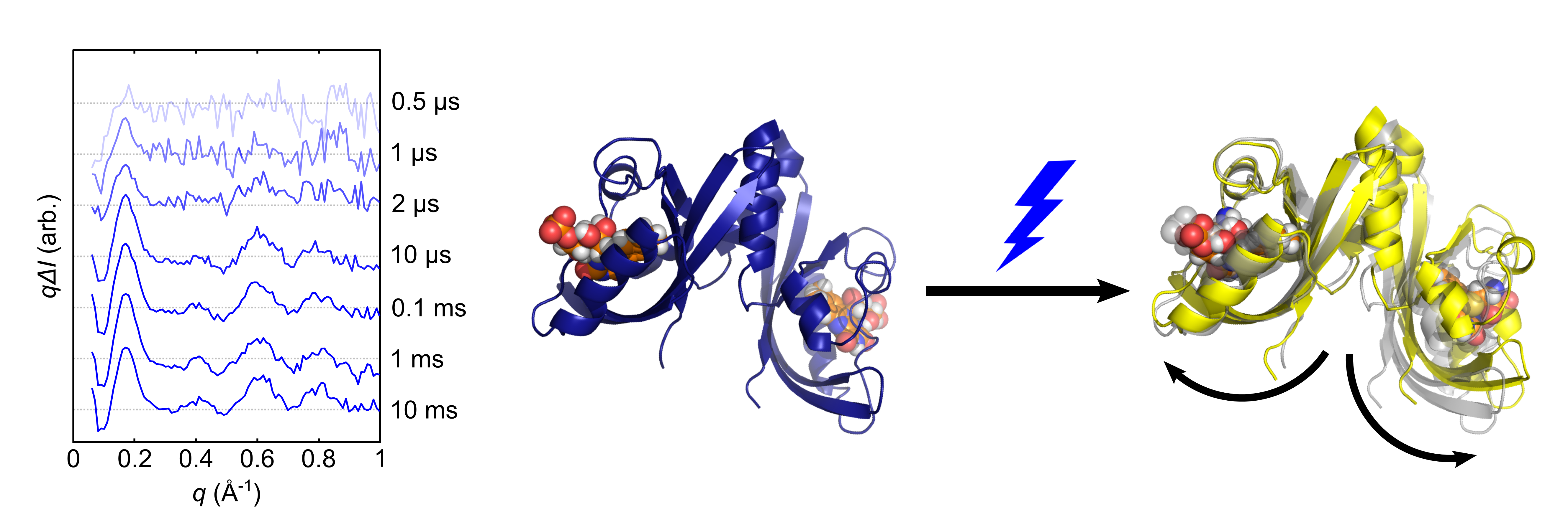
They first performed x-ray solution scattering of the complete YtvA LOV domain in the transition between dark and light. The Bio-CARS 14-ID-B x-ray beamline at the APS was used to record results between 0.5 microseconds and 10 milliseconds after a brief blue laser flash, and the Swiss Light Source (SLS) was used to record the data at times greater than 100 milliseconds. These resources provided the ability to record data at time scales compatible with the timing of the structural transition of the protein (Fig. 1). LOV is known to transform very quickly from a dark-adapted state through two photochemical intermediates before a bond is formed that stabilizes the state which transmits the downstream signal. X-ray solution scattering was able to capture this transition and provided data for comparison to molecular models of possible structural rearrangements.
Molecular dynamic simulations yielded approximately 50,000 possible dark- and light-state structures of the protein that were compared to the x-ray scattering data, thereby providing a clearer picture of the global structural changes that occur in the dark-to-light transition. The data showed that the two monomers of YtvA separate by about 3 Å in the dark-to-light transition (Fig. 1). The separation of the monomers occurs at the interface with the domain responsible for biochemical output, consistent with its role in signal transmission.
The helical domain, which was not present in the previous crystal structure, acts as a hinge allowing the monomers to move away from each other by about 6 degrees, levering the domains apart in a manner that suggests that this structural change is robust enough to be exploited for protein engineering projects.
The team has already extended this investigation to a blue-light-regulated histidine kinase using x-ray solution scattering recorded at the Bio-CARS beamline. Indeed, the solution x-ray scattering data provide a structural rationale for recent findings on a system for blue-light-regulated gene expression, thus facilitating the engineering of enhanced light-gated protein actuators. — Sandy Field
See: Oskar Berntsson1,7, Ralph P. Diensthuber2,7, Matthijs R. Panman1, Alexander Björling1, Ashley J. Hughes1, Léocadie Henry1, Stephan Niebling1, Gemma Newby3, Marianne Liebi4, Andreas Menzel4, Robert Henning5, Irina Kosheleva5, Andreas Möglich2*, and Sebastian Westenhoff1,8**, “Time-Resolved X-Ray Solution Scattering Reveals the Structural Photoactivation of a Light-Oxygen-Voltage Photoreceptor,” Structure 25, 933 (June 6, 2017). DOI: 10.1016/j.str.2017.04.006
Author affiliations: 1University of Gothenburg, 2Humboldt-Universität zu Berlin, 3European Synchrotron Radiation Facility, 4Paul Scherrer Institut, 5The University of Chicago
Correspondence: *[email protected],**[email protected]
S.W. acknowledges funding from the Swedish Foundation for International Cooperation in Research and Higher Education, the European Research Council, and the Foundation of Strategic Research, Sweden. A. Möglich acknowledges support from Deutsche Forschungsgemeinschaft through DFG grant MO2192/3-1 and a Sofja-Kovalevskaya Award by Alexander-von-Humboldt Foundation. Use of BioCARS was also supported by the NIGMS/NIH under grant number R24GM111072. The time-resolved setup at Bio-CARS was funded in part through a collaboration with Philip Anfinrud (NIH/NIDDK). This research used resources of the Advanced Photon Source, a U.S. DOE Office of Science User Facility operated for the DOE Office of Science by Argonne National Laboratory under Contract No. DE-AC02- 06CH11357.
Argonne National Laboratory seeks solutions to pressing national problems in science and technology. The nation's first national laboratory, Argonne conducts leading-edge basic and applied scientific research in virtually every scientific discipline. Argonne researchers work closely with researchers from hundreds of companies, universities, and federal, state and municipal agencies to help them solve their specific problems, advance America's scientific leadership and prepare the nation for a better future. With employees from more than 60 nations, Argonne is managed by UChicago Argonne, LLC for the U.S. Department of Energy's Office of Science.
The U.S. Department of Energy's Office of Science is the single largest supporter of basic research in the physical sciences in the United States and is working to address some of the most pressing challenges of our time. For more information, visit the Office of Science website.