
Manganese (Mn) acts as a cofactor to a variety of proteins necessary for proper bodily development and function. However, an overabundance of manganese in the brain can result in manganism, a neurological condition resembling Parkinson’s disease. Learning more about exactly which populations of neurons are affected by manganese accumulation is challenging due to the limitations of current imaging techniques. To overcome these limitations, researchers identified dopaminergic cells in rat brains using a retrograde fluorescent label and then performed x-ray fluorescence (XRF) microscopy at the U.S. Department of Energy’s Advanced Photon Source (APS) to determine the concentration and distribution of manganese in two regions of the substantia nigra. Rats chronically exposed to manganese exhibited a significant accumulation of manganese in both the substantia nigra pars compacta (>170%) and the substantia nigra pars reticulata (>163%), as compared to controls. Subcellular imaging of dopaminergic cells demonstrated that manganese is located primarily in the cytoplasm, where it would be available to interfere with dopamine production, regulation, and release. X-ray fluorescence data about the distribution of metals in the brain and their involvement in the development of neurodegenerative diseases can aid in the design of therapies.
Manganese is required for normal body functions and its dietary intake is regulated by gut absorption. Exposure to Mn, which is also critical to the production of steel, via inhalation or injection can increase the accumulation of the metal ion in the brain. This accumulation can result in a condition called manganism that shares many symptoms—tremors, stiffness, and slow movements—with Parkinson’s disease, a movement disorder resulting from the loss of dopaminergic (DA) neurons within the substantia nigra pars compacta (SNc). Cases of manganism have been reported in miners, welders, smelters, and abusers of ephedrine and methcathinone.
Direct observation of the overabundance of manganese in SNc could establish the biological basis underlying manganism. Cell culture experiments have determined bulk manganese concentrations, but have failed to model the effects of the blood-brain barrier on manganese access to the brain and the distribution of manganese among different brain structures. Magnetic resonance imaging (MRI) has been used to observe manganese distribution in different regions of the living brain. However, current MRI resolution falls far short of single-cell imaging. Conversely, techniques such as secondary ion mass spectroscopy that can provide subcellular resolution require ultra-thin samples and cannot be used for large-area imaging and therefore have not been used to localize manganese concentrations in the brain.
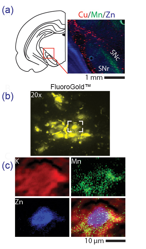
X-ray fluorescence microscopy—the emission of fluorescent x-rays from a material that has been bombarded with high-energy x-rays—presents an alternative approach. This technique permits the use of thick slices of brain tissue for manganese quantification with a wide range of possible spatial resolutions. In a series of experiments, researchers from Purdue University first used the in vivo retrograde tracer FluoroGold™ to fluorescently label dopaminergic cells of the SNc in rat brain tissue samples. They subsequently performed in situ tissue-level XRF scans (pixel size, 25 μm) of the SNc at the NSLS-II/X-ray Science Division [XSD]) 8-BM-B x-ray beamline and the Biophysics Collaborative Access Team 18-ID-D x-ray beamline, both at the Argonne APS, an Office of Science user facility. Additional cellular-level scans (pixel size, 0.30 μm) were performed at the XSD 2-ID-D and 2-ID-E beamlines, also at the APS.
A significant change in the manganese content of the SNc (>170%) was observed in manganese-treated groups as compared to control animals. Manganese was also significantly elevated in the neighboring substantia nigra pars reticulata region (>163%). Intracellular manganese concentrations in SNc ranged between 40–200 μM; concentrations as low as 100 μM have been observed to cause cell death in vitro. The plasma manganese concentration in rats after 30 days of manganese exposure was 11–36 μg/L, while manganese concentration in untreated animals was 3.5–5.6 μg/L. For reference, a recent study of manganese-poisoned welders who developed manganism had levels between 8.2–36 μg/L.
The researchers used strong zinc and potassium signals picked up in the XRF images to identify the location of SNc cell nuclei and the outlines of cell bodies, respectively (Fig. 1). They observed that manganese did not aggregate near the nucleus or the mitochondria as had been previously reported. Rather, manganese is heterogeneously distributed throughout the cell body, suggesting it is available to interfere with various structures controlling dopamine production, regulation, and release. Quantifying the accumulation of manganese in dopaminergic cells of the SNc may help clarify the relationship between manganese and the loss of motor skills associated with manganism.
— Chris Palmer
See: Gregory Robison, Brendan Sullivan, Jason R. Cannon, and Yulia Pushkar*, “Identification of dopaminergic neurons of the substantia nigra pars compacta as a target of manganese accumulation,” Metallomics 7(5), 748 (May 2015). DOI: 10.1039/c5mt00023h
Author affiliation: Purdue University
Correspondence: *[email protected]
This work was supported by National Institutes of Health/National Institute of Environmental Health Sciences grants R01 ES008146-14 (Y.P.) and R00ES019879 (J.R.C.) and by the Purdue University Research Foundation. This research used resources of the Advanced Photon Source, a U.S. Department of Energy Office of Science User Facility operated for the U.S. Department of Energy Office of Science by Argonne National Laboratory under Contract No. DE-AC02-06CH11357.
Argonne National Laboratory seeks solutions to pressing national problems in science and technology. The nation’s first national laboratory, Argonne conducts leading-edge basic and applied scientific research in virtually every scientific discipline. Argonne researchers work closely with researchers from hundreds of companies, universities, and federal, state and municipal agencies to help them solve their specific problems, advance America’s scientific leadership and prepare the nation for a better future. With employees from more than 60 nations, Argonne is managed by UChicago Argonne, LLC for the U.S. Department of Energy’s Office of Science.
The U.S. Department of Energy’s Office of Science is the single largest supporter of basic research in the physical sciences in the United States and is working to address some of the most pressing challenges of our time. For more information, visit the Office of Science website.