
Peptide amphiphiles (PAs) are an emerging class of molecules that can be designed for novel therapies in advanced medicine. They are designed with structural regions that allow them to spontaneously assemble into large complex structures like nanofibers (fibers with diameters of approximately 10 nanometers). Researchers in this study investigated how positively charged PAs interact with cells when water-hating properties and hydrogen bonding (a force that holds the nanofibers together) are altered. Using the BioCARS beamline 14-BM-C at the U.S. Department of Energy Office of Science's Advanced Photon Source (APS) to collect data, they evaluated forces within the biological assemblies. Their research, published in Nature Communications, shows that when bonding between molecules is weak, it allows them to rapidly associate with lipids in the cell membrane, leading to cell breakdown and death. In contrast, stronger bonds between molecules favor cell survival. These findings will be important in guiding future work to design biological materials for varied therapeutic use.
Peptides that are amphiphilic contain both water-loving (hydrophilic) and water-hating (hydrophobic) structural regions. Many of these peptides that exist in nature have positive charge (cationic) in the hydrophilic region, which contributes to various functions such as self-defense. It is their positively charged and water-hating regions that allow the peptides to associate so strongly with cell membranes that they can even lead to peptide breakdown. There are two classes of these peptides: cell penetrating peptides and natural host defense peptides, and both have proven useful in many therapeutic areas, including cancer therapy, antimicrobials, and delivery of genes or drugs.
Self-assembling peptides are a class of designer peptide-based molecules that make use of interactions between molecules that cause them to spontaneously assemble into large complex molecular structures of different shapes. Interest in these peptides continues to increase due to their potential for applications in the biomedical field, where they serve as building blocks for numerous materials and devices. This technology aims to mimic what happens in nature, basically using forces and bonds between molecules to produce ordered biological assemblies of building blocks with biochemical functions. One example of this involves nanofibers that mimic components of the mammalian extracellular matrix.
PAs are a type of self-assembling peptide. Some studies have shown that changing the amino acid sequence and degree of hydrogen bonding in PAs can significantly alter their shape and ability to assemble into three-dimensional networks. Other studies have also shown that changes in hydrogen bonding and electrostatic forces in these biological assemblies affects the strength of the materials they form, and their ability to signal cells. However, the interactions between cell membranes and these materials are not well understood. With this in mind, the researchers from Northwestern University aimed to investigate how hydrogen bonding, the presence of structural water-hating regions, and the charge of PAs affect how they interact with cells.
The researchers used data from wide-angle x-ray diffraction studies performed at the 14-BMC beamline at the Argonne APS, and showed that forces between molecules, particularly hydrogen bonding, influence the cell's ability to survive. They determined that differences in attractive forces in biological assemblies are the key in determining cell membrane stability, and therefore whether the cell dies or survives.
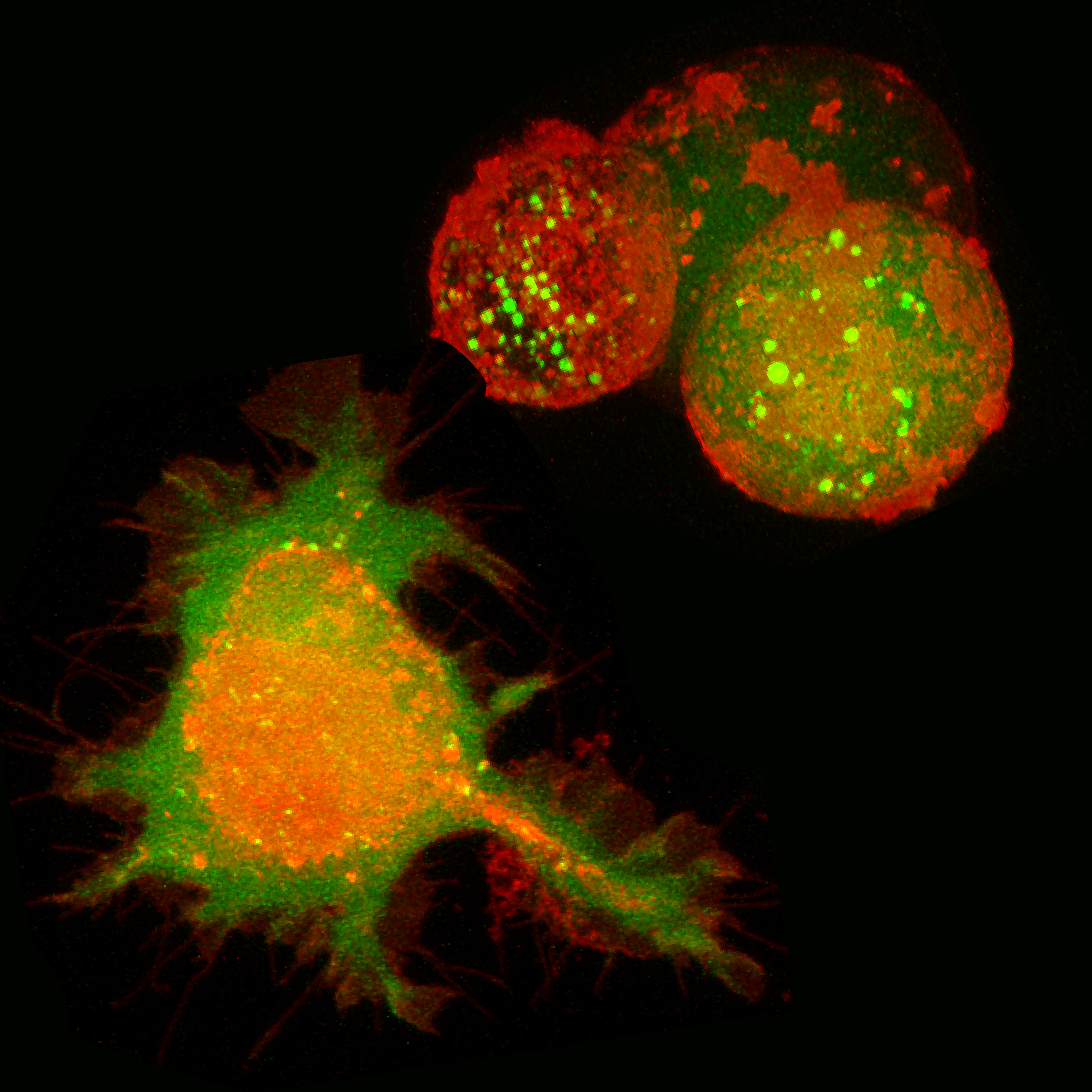
The most significant finding was in relation to changes in hydrogen bonding among molecules. The researchers showed that, when biological assemblies were fortified by strong hydrogen bond forces between molecules, cell survival was favored. However, when bonds between molecules were weak, biological assemblies were able to integrate into, and break down, the lipid layer of the cell membrane, and thereby stimulate cell death (see the figure). They also demonstrated that both the water-hating region and the positive charge of the PA were required to promote cell death.
"Borrowing nature's design principles, we can create materials that either support cell survival or cause cell death by simply changing how cohesive the material is," said Northwestern's Christina J. Newcomb, lead author of the Nature Communications paper.
— Nicola Parry
See: Christina J. Newcomb, Shantanu Sur, Julia H. Ortony, One-Sun Lee, John B. Matson, Job Boekhoven, Jeong Min Yu, George C. Schatz, and Samuel I. Stupp*, "Cell death versus cell survival instructed by supramolecular cohesion of nanostructures," Nat. Commun. 5, 3321(2014). DOI: 10.1038/ncomms4321
Author affiliation: Northwestern University
Correspondence: *[email protected]
This work was supported by the National Institutes of Health, National Institute of Dental and Craniofacial Research 2R01 DE015920-06, and the National Institute of Biomedical Imaging and Bioengineering 2R01EB003806-06A2. J.B. was supported by a Rubicon grant from the Netherlands Organisation for Scientific Research (NWO), and both O.-S.L. and G.C.S. were supported by National Science Foundation grant CHE-1147335. Use of BioCARS was supported by the National Institute of General Medical Sciences of the National Institutes of Health under grant number R24GM111072. Use of the Advanced Photon Source was supported by the U.S. Department of Energy, Basic Energy Sciences, Office of Science, under Contract No. DE-AC02-06CH11357.